Human embryo implantation recorded in real time for the first time
On Aug. 15, 2025, researchers at the Institute for Bioengineering of Catalonia (IBEC) in collaboration with the Dexeus University Hospital have captured unparalleled images of a human embryo implanting. This is the first time that the process has been recorded in real time and in 3D.
Failure of the implantation process in the uterus is one of the main causes of infertility, accounting for 60% of spontaneous abortions. Until now, it had not been possible to observe this process in humans in real time, and the limited available information came from still images taken at specific moments during the process.
‘We have observed that human embryos burrow into the uterus, exerting considerable force during the process. These forces are necessary because the embryos must be able to invade the uterine tissue, becoming completely integrated with it. It is a surprisingly invasive process. Although it is known that many women experience abdominal pain and slight bleeding during implantation, the process itself had never been observed before,” explains Samuel Ojosnegros, principal investigator of the IBEC’s Bioengineering for Reproductive Health group and leader of the study.
To advance during implantation, the embryo releases enzymes that break down the surrounding tissue. However, it is also known that force is required in order to penetrate the underlying layers of the uterus. This fibrous tissue is filled with collagen, a rigid protein that also forms tendons and cartilage. “The embryo opens a path through this structure and begins to form specialised tissues that connect to the mother’s blood vessels in order to feed,” adds Ojosnegros.
The research team’s results reveal that human embryos exert traction forces on their environment, remodelling it. Improving our understanding of the implantation process could have a significant impact on fertility rates, embryo quality and the time taken to conceive through assisted reproduction.
Tags:
Source: American Association for the Advancement of Science
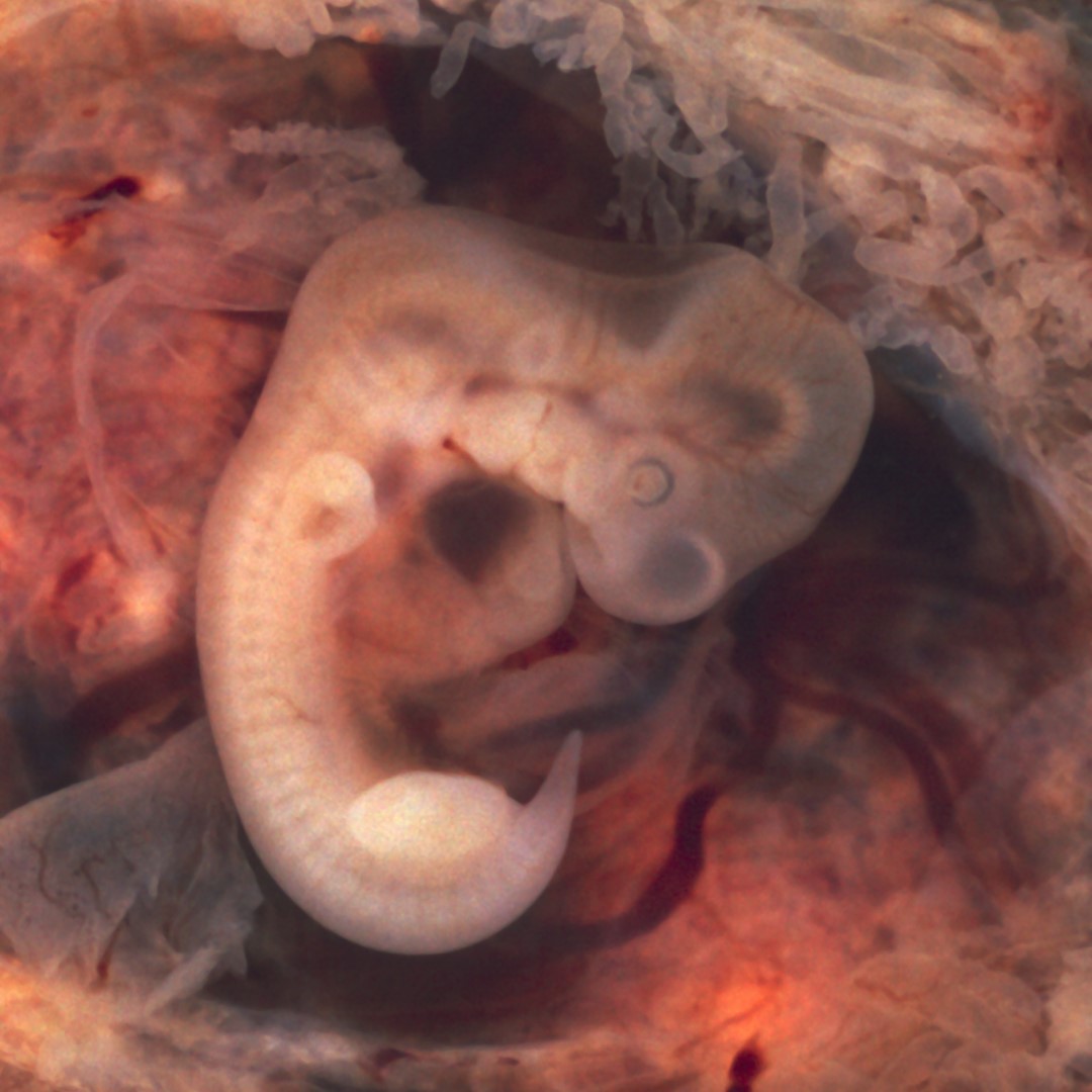
Credit: Photo: Photo of an opened oviduct with an ectopic pregnancy features a spectacularly well preserved 10-millimeter embryo (7th week of pregnancy). Courtesy: Ed Uthman, Flickr.